

Understanding thrombosis
Thrombosis is the formation of a clot in either an artery or a vein. The clot can slow and even stop blood flow. When a piece of the clot breaks off and travels through the circulation, it is referred to as an embolism.
Venous thromboembolism
Venous thromboembolism (VTE) is a blood clot that forms in a vein and manifests as deep vein thrombosis or pulmonary embolism.
"There are approximately 10 million cases of VTE annually, with up to 60% of cases occurring during or after hospitalization. In the U.S., there are 100,000—300,000 VTE-related deaths annually, with Europe recording 544,000 annually."

What is a pulmonary embolism?
Pulmonary embolism (PE) is a condition in which one or more arteries in the lung become blocked by a blood clot.
What is deep vein thrombosis?
Deep vein thrombosis (DVT) is a blood clot that forms in a vein deep in the body, occurring when blood thickens and clumps together. Most deep vein blood clots occur in the lower leg or thigh, but can also occur in other parts of the body.2


Slideshow: What do the symptoms of VTE look like?
The signs and symptoms of VTE are nonspecific and consistent with a variety of other conditions ranging from acute myocardial infarction and heart failure to pneumonia.

Why is early diagnosis important?
Properly assessed PE and DVT results can be a matter of life and death. Approximately 10% of diagnosed acute PE patients die within 60 minutes, and 80% of cases are not diagnosed until autopsy. PE is so common and lethal that the diagnosis should be sought actively in every patient who presents with any chest symptoms that cannot be proven to have another cause.*

How to diagnose PE
Diagnosing PE starts with a complete patient history and findings from a physical exam. Radiological procedures and laboratory tests provide important information when assessing patients suspected of VTE. In particular, the D-dimer test has shown to be extremely valuable in the diagnostic process for VTE.
Diagnostic tests
The D-dimer assay is a useful diagnostic aid for physicians treating patients with suspected thromboembolic disease, which can aid in determining whether further tests, including imaging, are needed. D-dimer testing is very cost-effective, requires a simple blood draw in order to perform the test, and provides results in just minutes.

Radiological tests
From a radiological perspective, pulmonary angiography is now the standard for the diagnosis of PE, but other techniques are also used.
Multidetector computed tomography angiography (CTA) is the initial imaging modality of choice for stable patients with suspected pulmonary embolism. The American College of Radiology (ACR) considers chest CTA to be the current standard of care for the detection of pulmonary embolism in these patients. CT scanning shows emboli directly, but it is noninvasive and less costly than pulmonary angiography.
Improving access to care with lab-quality solutions
Incorporating D-dimer testing into diagnostic algorithms for DVT and/or PE can be helpful in streamlining patient care. For patients with a low or moderate pretest probability score, a negative D-dimer test allows clinicians to exclude PE. This means clinicians can focus on identifying alternate causes of the patient’s symptoms or discharge them, if appropriate.
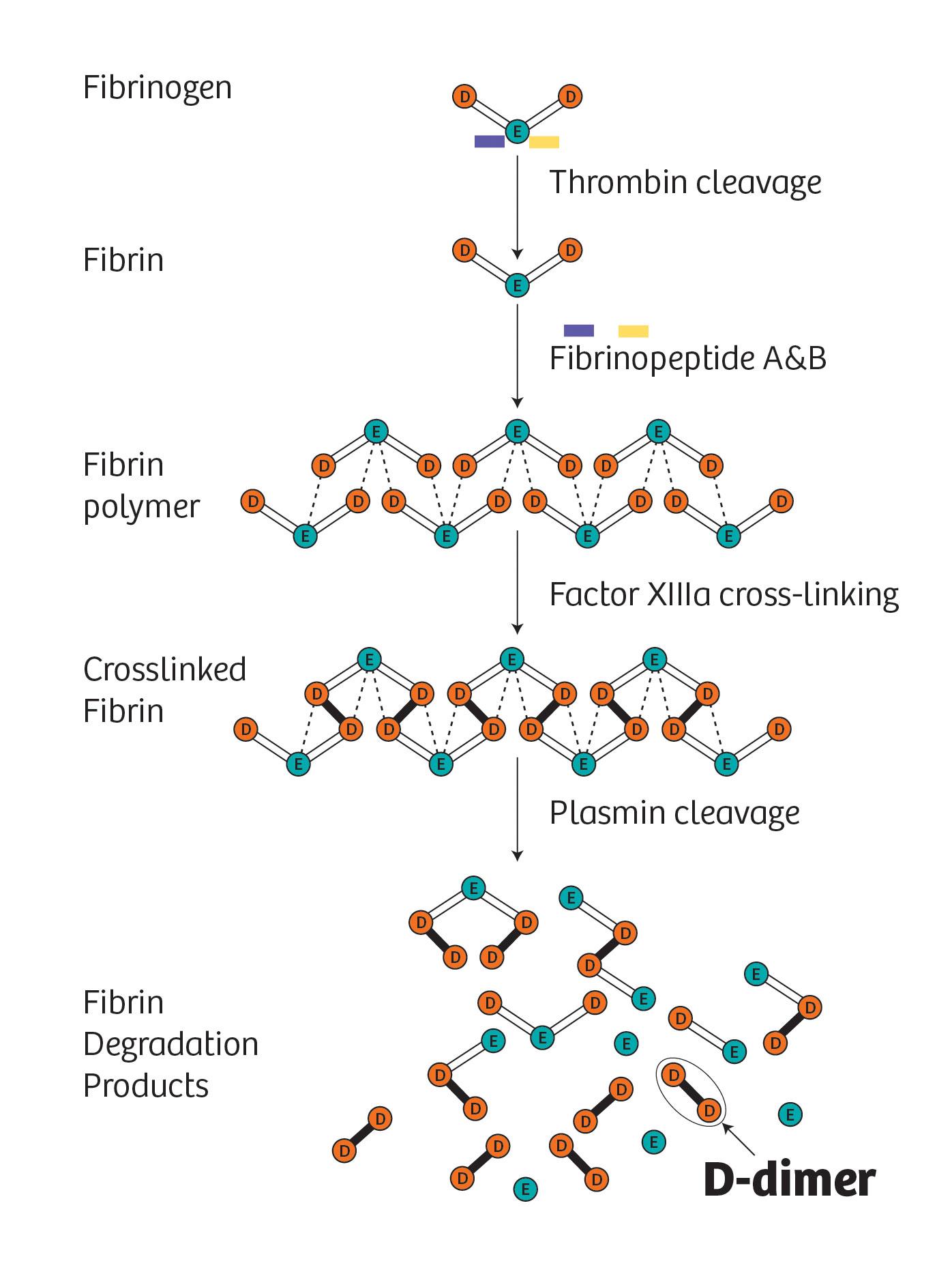
What is D-dimer?
D-dimer is a protein fragment produced when blood clots dissolve in the body. It is typically undetectable unless the body is forming and breaking down blood clots, at which point its level within the blood can rise. The D-dimer assay test detects D-dimer in the blood.
How Siemens Healthineers offers comprehensive cardiac testing
Our cardiac solutions help drive early diagnosis and treatment to improve clinical outcomes.
